DESCRIPTION
PHOSPHOLIPIDS are a class of lipids which constitute a major component of cell membranes and play important roles in signal transduction. Most phospholipids contain one diglyceride, a phosphate group, and one choline. BioAssay Systems' method provides a simple, direct and high-throughput assay for measuring choline-containing phospholipids in biological samples. In this assay, phospholipids (such as lecithin, lysolecithin and sphingomyelin) are enzymatically hydrolyzed to choline which is determined using choline oxidase and a H2O2 specific dye. The optical density of the pink colored product at 570 nm or fluorescence intensity (530/585 nm) is directly proportional to the phospholipid concentration in the sample.
KEY FEATURES
Sensitive. Use 20 µL samples. Linear detection range: colorimetric assay 3 - 200 µM, fluorimetric assay 0.6 - 20 µM phospholipid.
APPLICATIONS
Assays: phospholipid in biological samples such as serum and nonEDTA plasma.
Drug Discovery/Pharmacology: effects of drugs on choline-containing phospholipid metabolism.
KIT CONTENTS
Assay Buffer: 10 mL PLD Enzyme: 120 µL
Enzyme Mix: Dried Dye Reagent: 120 µL
Standard: 400 µL 2 mM phosphatidylcholine
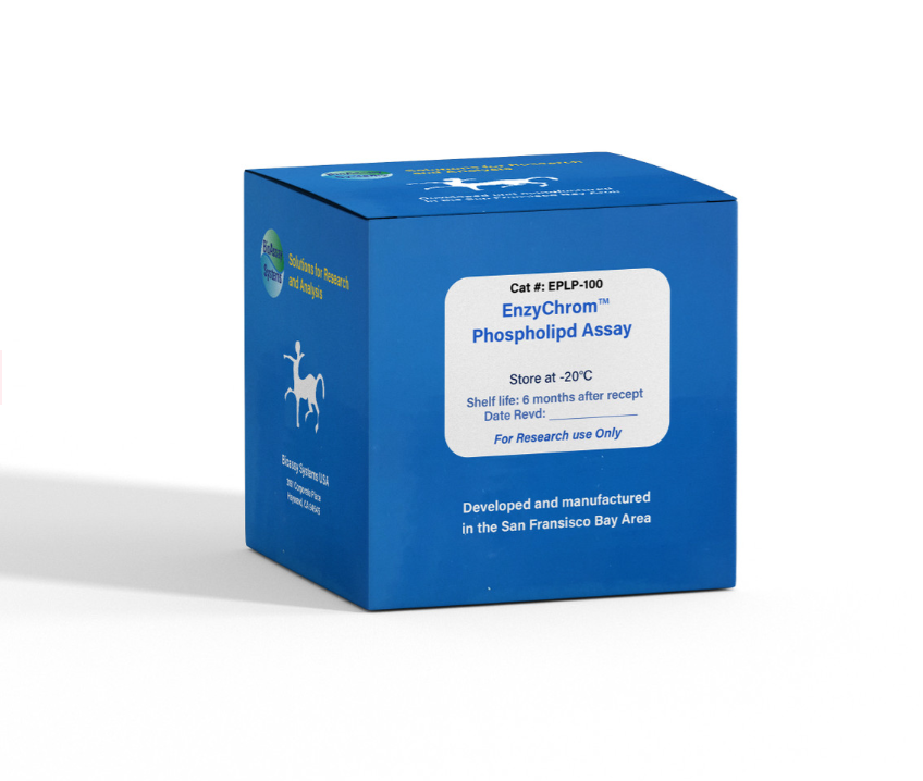
Storage conditions. The kit is shipped on ice. Store all components at -20°C. Shelf life of six months after receipt.
Precautions: reagents are for research use only. Normal precautions for laboratory reagents should be exercised while using the reagents. Please refer to Material Safety Data Sheet for detailed information.
COLORIMETRIC ASSAY
Liquid samples such as serum and plasma can be assayed directly. Solid samples can be homogenized in the assay buffer.
Note: SH-containing reagents (e.g. β–mercaptoethanol, dithiothreitol, > 5 µM), sodium azide, EDTA, and sodium dodecyl sulfate are known to interfere in this assay and should be avoided in sample preparation.
1. Equilibrate all components to room temperature. Briefly centrifuge the tubes before opening. Keep thawed tubes on ice during assay. Reconstitute Enzyme Mix with 120 µL Assay Buffer. Reconstituted Enzyme Mix is stable for 1 month when stored at -20˚C. Note: a yellow precipitate may form after thawing reconstituted Enzyme Mix. If a precipitate forms, pellet it by centrifuging for 2 min at 14000 rpm and use the clear supernatant.
2. Standards: mix 24 µL 2 mM Standard with 216 µL dH2O (final 200 µM). Dilute standard in dH2O as follows
Transfer 20 µL diluted standards into separate wells of a clear flatbottom 96-well plate.
Samples: transfer 20 µL of each sample into separate wells of the plate.
Note: if a sample is known to contain choline, prepare an extra sample blank well with 20 µL of the sample.
3. Color reaction. Prepare enough Working Reagent by mixing, for each well, 85 µL Assay Buffer, 1 µL PLD Enzyme, 1 µL Enzyme Mix and 1 µL Dye Reagent. Add 80 µL Working Reagent to each well. For samples that contain choline, prepare a blank control reagent with no PLD Enzyme (i.e., 85 µL Assay Buffer, 1 µL Enzyme Mix and 1 µL Dye Reagent). Add 80 µL of the Control Reagent to the Sample Blank well.
Tap plate to mix. Incubate 30 min at room temperature. Note: if precipitation occurs with certain samples, carry out the reaction in centrifuge tubes. After the 30 min incubation, centrifuge 5 min at 14,000 rpm. Transfer the supernatant into the wells for OD reading.
4. Read optical density at 570 nm (550-585 nm).
FLUORIMETRIC ASSAY
The fluorimetric assay procedure is similar to the colorimetric procedure except that (1) 0, 6, 12 and 20 µM phospholipid standards and (2) a black 96-well plate are used. Read fluorescence intensity at λex = 530 nm and λ em = 585 nm.
Note: if the calculated phospholipid concentration of a sample is higher than 200 µM in the Colorimetric Assay or 20 µM in the Fluorimetric Assay, dilute sample in 0.5% Triton X-100 and repeat the assay. Multiply result by the dilution factor n.
CALCULATION
Subtract blank value (#4) from the standard values and plot the ∆OD or ∆F against standard concentrations. Determine the slope and calculate the phospholipid concentration of Sample,
RSAMPLE and RBLANK are optical density or fluorescence intensity readings of the Sample and H2O Blank (or Sample Blank if sample contains choline), respectively. n is the sample dilution factor.
MATERIALS REQUIRED, BUT NOT PROVIDED
Pipetting devices, centrifuge tubes, clear flat-bottom uncoated 96-well plates, optical density plate reader; black flat-bottom uncoated 96-well plates, fluorescence plate reader.
PUBLICATIONS
1. Dhamdhere, GR et al (2014). Drugging a stem cell compartment using Wnt3a protein as a therapeutic. PloS one 9.1: e83650.
2. Kim, SH et al. (2019). The early onset and persistent worsening pulmonary alveolar proteinosis in rats by indium oxide nanoparticles. Nanotoxicology: 1-11.
3. Dai, W et al. (2020). OGDHL silencing promotes hepatocellular carcinoma by reprogramming glutamine metabolism. Journal of Hepatology, 72(5): 909-923.
