DESCRIPTION
SUPEROXIDE DISMUTASES (SOD, EC1.15.1.1) are enzymes that catalyze the dismutation of superoxide into O2 and H2O2. They are an important antioxidant defense in all cells exposed to O2. There are three major families of superoxide dismutase: Cu/Zn, Fe/Mn, and the Ni type. Aberrant SOD activities have been linked to diseases such as amyotrophic lateral sclerosis, perinatal lethality, neural disorders and cancer.
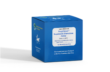
BioAssay Systems’ SOD assay provides a convenient colorimetric means for the quantitative determination of SOD enzyme activity in biological samples. In the assay, superoxide (O2 - ) is provided by xanthine oxidase (XO) catalyzed reaction. O2 - reacts with a WST-1 dye to form a colored product. SOD scavenges the O2 - thus less O2 - is available for the chromogenic reaction. The color intensity (OD440nm) is used to determine the SOD activity in a sample.
KEY FEATURES
Sensitive and accurate. Linear detection range of 0.05 - 3 U/mL SOD.
Convenient and high-throughput. Homogeneous "mix-incubate-measure" type assay. No wash and reagent transfer steps are involved. Can be readily automated on HTS liquid handling systems for processing thousands of samples per day.
APPLICATIONS
Determination of SOD in blood, cell, tissue and other biological samples.
KIT CONTENTS
Assay Buffer: 20 mL Diluent: 20 mL
SOD Enzyme: 120 µL XO Enzyme: 120 µL
Xanthine: 600 µL WST-1: 600 µL
Storage conditions: The kit is shipped on ice. Store all reagents at -20°C. Shelf life of 6 months after receipt.
Precautions: reagents are for research use only. Normal precautions for laboratory reagents should be exercised while using the reagents. Please refer to Material Safety Data Sheet for detailed information.
ASSAY PROCEDURE
Note: If not assayed immediately, samples can be stored at -80°C for one month. All samples can be diluted in 50 mM potassium phosphate, pH 7.4.
1. Tissue samples. Perfuse tissue with cold PBS to remove any red blood cells. Homogenize tissue at 5 mL/g in cold lysis buffer (50mM potassium phosphate, 0.1 mM EDTA, 0.5% Triton X-100). Centrifuge at 12,000g for 5 minutes at 4°C. Use supernatant for total SOD assay.
2. Cell samples. Suspension cells: Centrifuge 1-2 x 10⁶ cells at 800g for 2 minutes and discard supernatant. Wash cells with cold PBS, centrifuge, and discard the supernatant. Resuspend cells in 0.5 mL of cold lysis Buffer. After 10 min on ice, centrifuge at 12,000g for 5 min. Use supernatant for total SOD assay. Adherent cells: Wash 1-2 x 10⁶ cells cold PBS. Place dish on ice. Add 0.5 mL of cold lysis buffer. After 10 min on ice, collect cells/debris with a rubber policeman. Centrifuge the cell extract at 12,000g for 5 min. Use supernatant for total SOD assay. Note: if it is desired to determine cytosolic and mitochondrial SOD activities separately, tissue/cell samples can be prepared according to Mattiazzi et al (2002). JBC 277: 29626-33.
3. Blood samples: Collect serum, or plasma (heparin, citrate or EDTA) using standard protocols. The erythrocyte pellet can be lysed in 5x volume of cold dH2O; centrifuge at 12,000g for 5 min to pellet the erythrocyte membranes. Dilute serum/plasma 1:5, red cell lysate 1:100 prior to SOD assay.
Prior to assay, bring all reagents to room temperature (25°C). The Xanthine reagent may appear to be turbid. Briefly vortext this tube before pipetting. Briefly centrifuge enzyme tubes, keep on ice during assay
1. Standards. Mix 8µL SOD Enzyme with 392µL Diluent to give 3U/mL SOD standard. Dilute standards as below.
Transfer 20 µL SOD standards to separate wells of a clear flat-bottom 96-well plate. Transfer 20 µL samples to separate wells.
2. Prepare enough Working Reagent for the standard and sample wells. For each well, mix 160 µL Assay Buffer, 5 µL Xanthine and 5 µL WST-1. Transfer 160 µL Working Reagent to each well and tap plate to mix. For each well, dilute the XO Enzyme 1:20 in Diluent. Quickly add 20 µL diluted XO enzyme to each assay well (use of a multi-channel pipettor is recommended). Tap plate to mix.
3. Immediately read OD440nm (OD420-460nm) (ODo). Incubate for 60 min at room temperature (25°C) in the dark. Read OD440nm again (OD60).
CALCULATION
1. For each standard and sample well, calculate ∆OD60 = OD60 - ODo.
2. Calculate ∆∆OD = ∆ODstd8 - ∆OD for each standard and sample where ∆ODstd8 is the ∆OD for Standard # 8 (the standard with no SOD activity and highest possible absorbance).
3. Plot the Standard Curve ∆∆OD vs [SOD](U/mL). Use the ∆∆OD for sample to determine SOD activity of sample from the standard curve.
MATERIALS REQUIRED, BUT NOT PROVIDED
Pipetting devices, tissue homogenizer, centrifuge and tubes, clear flatbottom 96-well plates and plate reader.
PUBLICATIONS
1. Magana-Cerino, J. M., et al. (2020). Consumption of nixtamal from a new variety of hybrid blue maize ameliorates liver oxidative stress and inflammation in a high-fat diet rat model. Journal of Functional Foods. 72: 104075
2. Nawi, A., et al. (2020). Lipid peroxidation in the descending thoracic aorta of rats deprived of REM sleep using the inverted flowerpot technique. Experimental Physiology. 105(8): 1223-1231.
3. Sahin, T. D., et al. (2020). Infliximab prevents dysfunction of the vas
deferens by suppressing inflammation and oxidative stress in rats with
chronic stress. Life Sciences. 250: 117545.
